

Finding out you are pregnant brings a rush of emotions. If this is your first time, your first trimester ultrasound appointment can feel a little mysterious. What will they actually see? What do the measurements mean? What does all of it tell you about how your baby is doing?
This week by week guide covers exactly what shows up on early pregnancy ultrasounds so you can walk into that appointment feeling informed and ready.
A Quick Note on the Type of Ultrasound
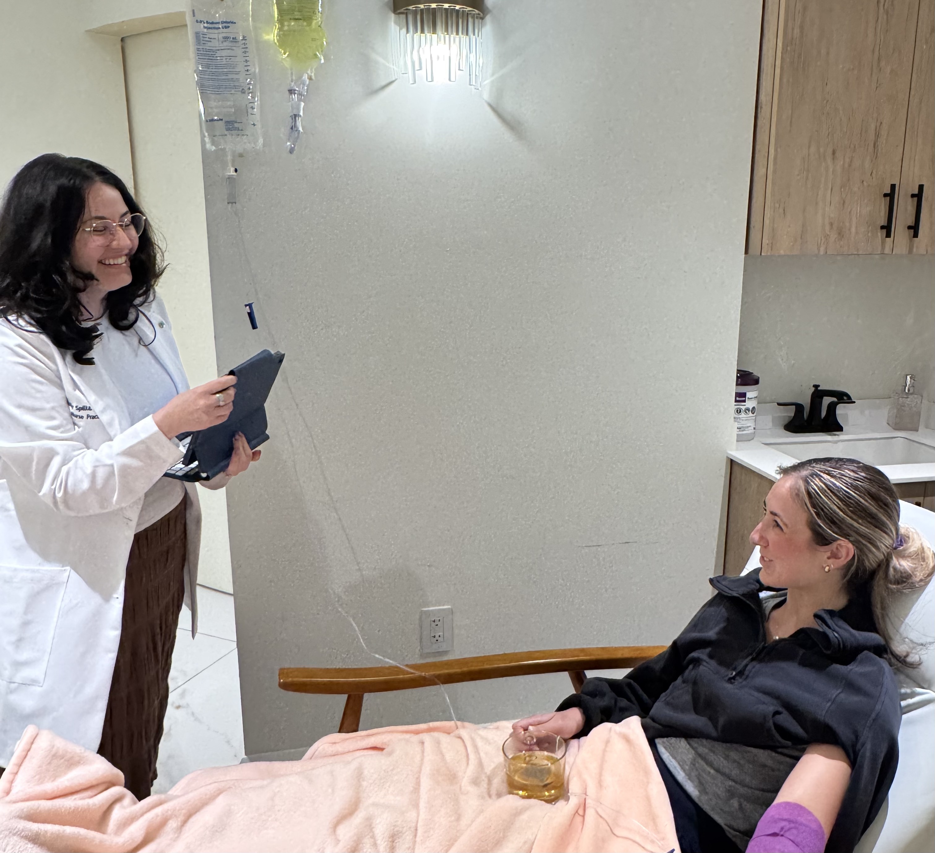
In early pregnancy, especially before 10 weeks, your provider will likely use a transvaginal ultrasound rather than the abdominal ultrasound you might be picturing. A small wand is gently placed inside the vagina to get a much closer view of the uterus. It sounds intimidating, but it is generally not painful, and it gives a significantly clearer picture than an external scan can at this stage of early pregnancy.
Week 4: Too Early to See Much, and That Is Normal
At four weeks, you have just missed your period and probably just taken a positive pregnancy test. Your uterus is still getting organized, and even on a transvaginal ultrasound, there is usually not much to see yet. The fertilized egg has implanted, but the structures that show up on an early pregnancy scan have not yet formed. If you have an ultrasound at this point and do not see much, do not worry. This is completely expected.
Week 5: The Gestational Sac Appears
This is usually the first week something becomes visible on your pregnancy ultrasound. A small round or oval shape called the gestational sac appears on screen. Think of it as the earliest home your embryo will grow inside. At this point, a provider might refer to it as a probable pregnancy, meaning everything looks like it should, but they will want to confirm more at a follow up scan.
Week 5.5: The Yolk Sac Shows Up
Nestled inside the gestational sac, a tiny circular structure called the yolk sac appears. It is about the size of a small pea and serves as an early nutrient source for the embryo before the placenta takes over. Seeing the yolk sac is a significant milestone. It confirms that the pregnancy is inside the uterus, which rules out an ectopic or tubal pregnancy. Your provider will likely confirm this as a definite intrauterine pregnancy at this point.
Week 6: Your Baby's First Heartbeat on Ultrasound
This is the appointment many parents describe as life changing. Around six weeks, the embryo becomes visible on a first trimester ultrasound for the very first time. It is a tiny structure next to the yolk sac, often measuring just a few millimeters. Alongside it comes cardiac activity. On the screen, it looks like a rapid flickering motion. It is not a fully formed heart yet, but it is real movement driven by electrical activity and the very beginning of your baby's heartbeat. Most providers will let you see and hear it.
Week 8: Everything Is Taking Shape
By eight weeks, the embryo is clearly visible, growing quickly, and starting to look more like a tiny person with each passing day. Your provider will begin measuring the crown rump length or CRL, which is the distance from the top of the head to the bottom of the spine. CRL measurement is the most accurate method for confirming how far along you are during an early pregnancy scan. Basic body structures are forming, and cardiac activity should be strong and steady.
Week 10: Growing Fast
Growth continues rapidly. By ten weeks, the embryo is roughly an inch long and early features including limb buds and facial profile are becoming more distinct. The heartbeat is robust. If your provider did not establish your due date at an earlier scan, this is still an excellent window for accurate first trimester pregnancy dating.
Week 12: The Big First Trimester Scan
Many OB practices schedule a detailed first trimester ultrasound at or around 12 weeks. By now, your baby looks unmistakably human. You can see the head, arms, legs, and face in profile. This is also when your provider will measure the nuchal translucency, which is a thin fluid filled space at the back of the baby's neck. This measurement, combined with a blood test, is part of first trimester screening for chromosomal differences including Down syndrome.
What Does Monitoring Closely Actually Mean?
If your provider mentions that a measurement looks borderline, or suggests coming back in a week for a follow up scan, try not to spiral. Early pregnancy ultrasound involves close monitoring precisely because the margins are tight and measurements can vary depending on the equipment, the angle, and who is reading the scan. Hearing that your provider wants to take another look next week is standard clinical practice and not necessarily a sign that something is wrong.
If there are genuine concerns, your provider will typically ask a second clinician to confirm before making any diagnosis. That is by design, and it is there to protect you.
How Your Due Date Is Determined from Ultrasound
Your first trimester ultrasound is actually more accurate than your last menstrual period for calculating your due date, especially if your cycles are irregular. The crown rump length measurement in early pregnancy can pin down your due date to within about five days. If your ultrasound dating differs by more than a week from what you calculated, your provider may adjust your estimated due date accordingly. Having the most accurate number possible is what matters most.
What to Bring to Your Ultrasound Appointment
Bring a support person if that feels right for you. Some providers ask you to arrive with a moderately full bladder for early scans, so check with your office in advance. Write down any questions you have beforehand so you will not forget them in the moment.
Your first trimester ultrasounds are as much about giving you information and reassurance as they are about clinical data. You are allowed to ask what you are looking at, what the measurements mean, and what comes next. That is exactly what these appointments are for.
We're here for you - from the start
We’re redefining maternal health by replacing the ER with a welcoming, evidence-based care environment that centers you: your body, your symptoms, your experience.
Schedule a visit
Compassionate support for every stage of pregnancy
Contact
Connect with our dedicated support team
Phone
Direct line for immediate healthcare guidance
Office
Women's health center in
130 7th Ave S NY, NY 10014
130 7th Ave S NY, NY 10014
Related blogs and articles
Migraines in Pregnancy: What Is Safe and When to Worry
Migraines in pregnancy are common and most are not dangerous. An NYC OB/GYN explains what is safe, the red flags, and same day care. Book today.

When Is My Fertile Window, and Did I Miss It?
Wondering when your fertile window is, and if you missed it? An NYC nurse midwife explains how to know, and how to confirm it. Same week appointments available.
Postpartum Care Beyond Six Weeks: What Your Body Is Actually Going Through
The 6-week visit is not the finish line. Here is what your body needs postpartum beyond six weeks and what real fourth trimester care covers.